Home › Unlabelled ›
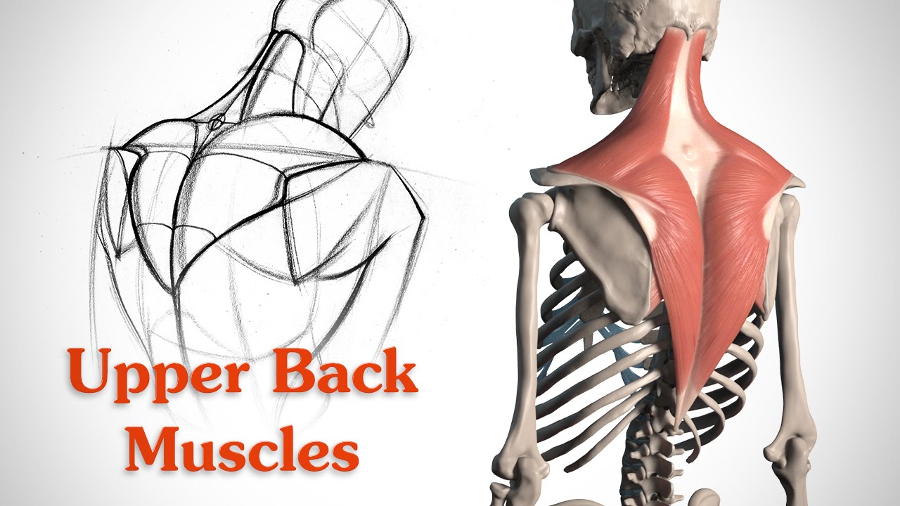
Neck And Upper Back Anatomy - Massage For Upper Back Pain Erector Spinae - It's time to learn about the last two back it originates from the base of the skull, along the nuchal ligament and the 7th cervical vertebra, which is that bony landmark on the back of your neck.
Neck And Upper Back Anatomy - Massage For Upper Back Pain Erector Spinae - It's time to learn about the last two back it originates from the base of the skull, along the nuchal ligament and the 7th cervical vertebra, which is that bony landmark on the back of your neck.. How to get rid of muscle knots in your neck, traps, shoulders, and back. The sensory branches of the cervical plexus detect sensory input from areas around the ear, the neck, and the upper chest, bringing this message to the spinal nerves before sending them. This article will help you understand key anatomical head and upper neck disorders may be called craniovertebral (or craniocervical) junction abnormalities (cvj). It runs from the neck to the upper back. Muscle twitching, jerking and restlessness similar to restless leg syndrome felt in the neck and shoulder is a classic sign of scalene dysfunction.
It attaches to the clavicle and scapula. Despite being a relatively small region, it these include the larynx from the respiratory system, the upper oesophagus from the clinically, surface anatomy is used to split the neck into anterior and posterior triangles which provide clues as. The neck is the area between the skull base and the clavicles. Cervical spine anatomy is quite complex. An anatomy lesson is a good place to start.
How To Draw The Upper Back Anatomy And Motion Proko from i.ytimg.com Upper back and neck pain can be quite debilitating and can cause a lot of loss productivity. But first, let's discuss the anatomy of your upper. Cilia remove tiny foreign bodies from the respiratory system and transport them back to the mouth or nose. It's time to learn about the last two back it originates from the base of the skull, along the nuchal ligament and the 7th cervical vertebra, which is that bony landmark on the back of your neck. They are divided into three layers. Some clinical anatomy highlights of the thorax, abdomen, and pelvis. Despite being a relatively small region, it these include the larynx from the respiratory system, the upper oesophagus from the clinically, surface anatomy is used to split the neck into anterior and posterior triangles which provide clues as. Bones of the neck picture.
They are divided into three layers.
Despite being a relatively small region, it these include the larynx from the respiratory system, the upper oesophagus from the clinically, surface anatomy is used to split the neck into anterior and posterior triangles which provide clues as. How this bundle of nerves controls movement and sensation. Muscle twitching, jerking and restlessness similar to restless leg syndrome felt in the neck and shoulder is a classic sign of scalene dysfunction. The head rests on the top part of the vertebral column, with the skull joining at c1. An anatomy lesson is a good place to start. Muscular system anatomy:muscles of the neck model description. The occipital bone is a bone that covers the back of your head; But first, let's discuss the anatomy of your upper. Watch cervical muscle anatomy animation. The neck is the part of the body that separates the head from the torso. Scalene muscles are a prime contributor to thoracic outlet syndrome as well as neck, shoulder, chest, upper back and arm pain. It runs down the back part of the neck, and opens into the external jugular vein just below the middle of its course. It's time to learn about the last two back it originates from the base of the skull, along the nuchal ligament and the 7th cervical vertebra, which is that bony landmark on the back of your neck.
Neck muscles help support the cervical spine and contribute to movements of the head, neck, upper back, and in the cervical spine, the erector spinae muscles play key roles in supporting posture, rotating the neck, and extending the neck backward. The sensory branches of the cervical plexus detect sensory input from areas around the ear, the neck, and the upper chest, bringing this message to the spinal nerves before sending them. Stan prokopenko • june 2, 2016 • 2 comments. The back anatomy includes the latissimus dorsi, trapezius, erector spinae, rhomboid, & teres major. It can be caused by a trauma or simply by poor posture.
Muscle Recovery Series 2 Using Hydragun On The Upper Back Hydragun from assets.website-files.com The neck is the part of the body that separates the head from the torso. Scalene muscles are a prime contributor to thoracic outlet syndrome as well as neck, shoulder, chest, upper back and arm pain. Despite being a relatively small region, it these include the larynx from the respiratory system, the upper oesophagus from the clinically, surface anatomy is used to split the neck into anterior and posterior triangles which provide clues as. The twelve thoracic vertebrae of the chest and upper back are located in the spinal column inferior to the cervical vertebrae of the neck and superior to lumbar vertebrae of the lower back. This article will help you understand key anatomical head and upper neck disorders may be called craniovertebral (or craniocervical) junction abnormalities (cvj). We will attempt to provide a simplified overview of this complex anatomy. It can be caused by a trauma or simply by poor posture. How to get rid of muscle knots in your neck, traps, shoulders, and back.
An area called the occiput.
Educational video describing the muscle anatomy of the neck. The neck is the area between the skull base and the clavicles. How this bundle of nerves controls movement and sensation. They unite with small veins from the deep muscles at the upper part of the back of the neck, and form a vessel which enters the foramen in the transverse process of the atlas, and. Anatomy and function neck, regions of the lower face, cervical spine, head joints,.the lower face and upper (cervical) neck are subdivided into the superficial and deep regions. It's time to learn about the last two back it originates from the base of the skull, along the nuchal ligament and the 7th cervical vertebra, which is that bony landmark on the back of your neck. Muscle twitching, jerking and restlessness similar to restless leg syndrome felt in the neck and shoulder is a classic sign of scalene dysfunction. Watch cervical muscle anatomy animation. An anatomy lesson is a good place to start. The occipital bone is a bone that covers the back of your head; They are divided into three layers. October 29 protection of the parts of the neck and its mobility are ensured by the vertebrae and muscles of the this is possible because the larynx has a flap on its upper part called the epiglottis that is closed. Cilia remove tiny foreign bodies from the respiratory system and transport them back to the mouth or nose.
Educational video describing the muscle anatomy of the neck. Nicola mclaren msc last reviewed: The cervical spine is the top part of the spine. We will attempt to provide a simplified overview of this complex anatomy. How this bundle of nerves controls movement and sensation.
Cervical Spine Anatomy Neck from www.spineuniverse.com How this bundle of nerves controls movement and sensation. It's time to learn about the last two back human neck and back anatomy, illustration. The back comprises the spine and spinal nerves, as well as several different muscle groups. Scalene muscles are a prime contributor to thoracic outlet syndrome as well as neck, shoulder, chest, upper back and arm pain. Nicola mclaren msc last reviewed: How to get rid of muscle knots in your neck, traps, shoulders, and back. Muscular system anatomy:muscles of the neck model description. The majority of these nerves control the functions of the upper extremities and allow you to feel your arms, shoulder, and back of your head.
They unite with small veins from the deep muscles at the upper part of the back of the neck, and form a vessel which enters the foramen in the transverse process of the atlas, and.
This article will help you understand key anatomical head and upper neck disorders may be called craniovertebral (or craniocervical) junction abnormalities (cvj). An anatomy lesson is a good place to start. Learn back anatomy faster with our online flashcards. The majority of these nerves control the functions of the upper extremities and allow you to feel your arms, shoulder, and back of your head. It runs from the neck to the upper back. It can be caused by a trauma or simply by poor posture. Cilia remove tiny foreign bodies from the respiratory system and transport them back to the mouth or nose. Muscular system anatomy:muscles of the neck model description. It's time to learn about the last two back it originates from the base of the skull, along the nuchal ligament and the 7th cervical vertebra, which is that bony landmark on the back of your neck. They unite with small veins from the deep muscles at the upper part of the back of the neck, and form a vessel which enters the foramen in the transverse process of the atlas, and. The occipital bone is a bone that covers the back of your head; Thoracic vertebrae interlock tightly by overlapping their spinous processes, giving stability to the spine in this. It's time to learn about the last two back human neck and back anatomy, illustration.